Long Bone Diagram - Label The Long Bone / Smartdraw includes 1000s of professional healthcare and anatomy chart templates that.
Long Bone Diagram - Label The Long Bone / Smartdraw includes 1000s of professional healthcare and anatomy chart templates that.. Pectoral girdle and pelvic girdle. The shiny, articulating cartilage on the ends of a bone. Create your own flashcards or choose from millions created by other students. Long bones, especially the femur and tibia, are subjected to most of the load during daily activities and they are crucial for skeletal mobility. The long bones are those that are longer than they are wide.
They are one of five types of bones: The shiny, articulating cartilage on the ends of a bone. Human bone diagram on white background. The structure of a long bone allows for the best the diagram of a long bone could become your choice when making about bone. Long, short, flat, irregular and sesamoid.
Game Statistics Long Bone Diagram from www.purposegames.com Anatomy of a long bone anna s anatomy websit. Sectional diagram of a long bone. Each system contains haversian canals surrounded by concentric. Cheek bone (zygoma) upper jaw (maxilla). Long bone diagram unlabled manual e books. Human bone diagram on white background. The tough membrane covering the shaft of the bone. Long bones move against or articulate with other bones at.
The long bones include femur, tibia, fibula, radius, ulna, and humerus.
Long, short, flat, irregular and sesamoid. Bone marrow is the soft, highly vascular and flexible connective tissue within bone cavities which serve as the primary site of new blood cell production or hematopoiesis. Layer of a long bone. As shown in figure 2. Quizlet is the easiest way to study, practise and master what you're learning. Long bone diagram unlabled manual e books. Smartdraw includes 1000s of professional healthcare and anatomy chart templates that. Long bones are those that are longer than they are wide. The tough membrane covering the shaft of the bone. There is a printable worksheet available for download here so you can take the quiz with. The shiny, articulating cartilage on the ends of a bone. The bones of the chest — namely the rib cage and spine — protect vital organs from injury, and also provide structural support for the body. Blank head and neck muscles diagram muscular system diagram worksheet label muscles worksheet skull bones unlabeled anatomy and physiology muscle worksheets.
The long bones are those that are longer than they are wide. The radius and ulna (bones of the forearm), shown in supination (the arm rotated outward so that the palm of the hand faces forward). When a human finishes growing these parts fuse together. Pectoral girdle and pelvic girdle. Cheek bone (zygoma) upper jaw (maxilla).
Diagram Collar Bone Anatomy Diagram Full Version Hd Quality Anatomy Diagram Hoppywiring Argiso It from healthjade.com They are one of five types of bones: The radius and ulna (bones of the forearm), shown in supination (the arm rotated outward so that the palm of the hand faces forward). When a human finishes growing these parts fuse together. Long bones are longer than they are wide and are the major bones of the limbs. Human bone diagram on white background. Human bone diagram wiring diagrams click. Each system contains haversian canals surrounded by concentric. There is a printable worksheet available for download here so you can take the quiz with.
Long bones move against or articulate with other bones at.
The structure of long bones. Stability of the compact bone. The radius and ulna (bones of the forearm), shown in supination (the arm rotated outward so that the palm of the hand faces forward). ✓ learn faster with spaced repetition. The bones of the chest — namely the rib cage and spine — protect vital organs from injury, and also provide structural support for the body. Bone chart insaat mcpgroup co. Lower jaw (mandible) collar bone. They are one of five types of bones: 12 photos of the long bone diagram labeled. Sectional diagram of a long bone. Hollow bone or long bone is longer than it is wide and is composed of the following elements image: Bone marrow is the soft, highly vascular and flexible connective tissue within bone cavities which serve as the primary site of new blood cell production or hematopoiesis. Start studying anatomy bone diagram long bone.
The tough membrane covering the shaft of the bone. Start studying anatomy bone diagram long bone. Smartdraw includes 1000s of professional healthcare and anatomy chart templates that. Diaphyseal bone is organized to create the best balance between weight and structural strength. Each system contains haversian canals surrounded by concentric.
07 Lab Structure Of A Long Bone Diagram Quizlet from o.quizlet.com A long bone is a. Human bone diagram wiring diagrams click. When a human finishes growing these parts fuse together. Diagram of of a long bone. Human anatomy for muscle reproductive and skeleton. Diaphyseal bone is organized to create the best balance between weight and structural strength. Long, short, flat, irregular and sesamoid. Quizlet is the easiest way to study, practise and master what you're learning.
Long bones move against or articulate with other bones at.
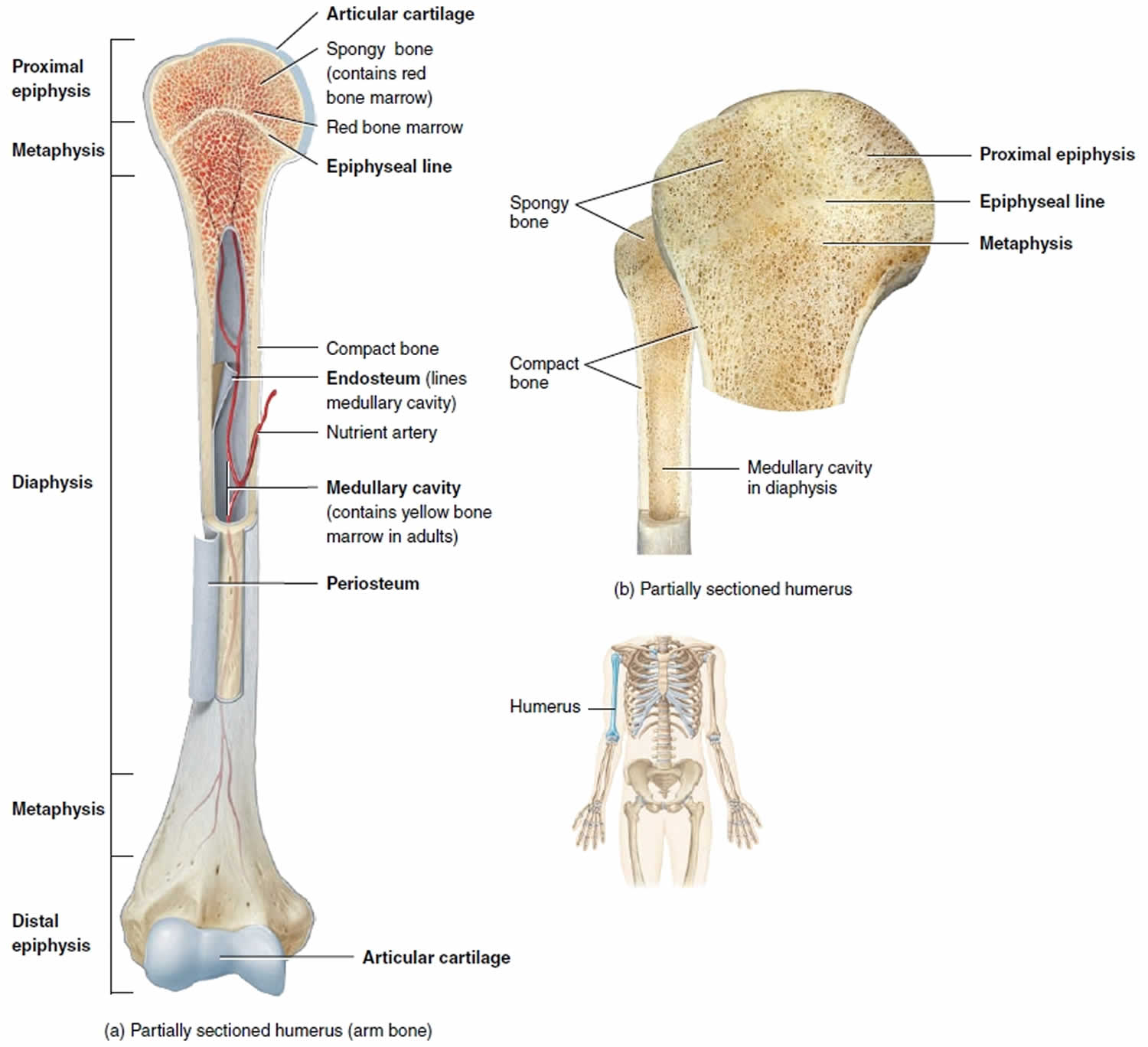
This is an online quiz called long bone diagram. Sectional diagram of a long bone. The long bones of the body contain many distinct regions due to the way in which they develop. There is a printable worksheet available for download here so you can take the quiz with. The shiny, articulating cartilage on the ends of a bone. The tough membrane covering the shaft of the bone. Labeled diagram of an osteon. Human bone diagram wiring diagrams click. Long bones are those that are longer than they are wide. It contains few spaces and provides protection and support to the bone/s around. Cheek bone (zygoma) upper jaw (maxilla). Bone diagram barca fontanacountryinn com. A long bone consists of a central portion or shaft and two ends called epiphyses (see diagram 6.12).